Chap5 The Big Wave is Coming, Drastic Changes in Depression and Schizophrenia Diagnosis
Big wave
Dramatic changes in the diagnosis of depression and schizophrenia will begin at the end of this year, 2021. A big wave of change is coming to us.Writing this in May 2021. The dramatic changes are more than seven months away, and they haven’t even happened yet. But I’m saying that will be the start of dramatic changes in late 2021.
It looks like I am a prophet who can see the future, but I am not a prophet.
I’m describing the facts that will inevitably happen, and I’m predicting a chain reaction of events that will start from those planned facts, and I’m predicting the big wave of the changes.
What are the dramatic changes in depression and schizophrenia diagnosis?
Diagnosis
In a nutshell, the diagnosis of depression and schizophrenia will change from a subjective diagnosis to an objective one.It changes from a literary diagnosis that listens to the patient’s mood to a scientific diagnosis that focuses on the results of brain imaging. The size of the brain injury will indicate the severity of the symptoms and the recovery status.
Objective judgment of the effect of treatment
The effect of the treatment can be judged objectively.Brain imaging results show the change in the size of the amygdala injury in depression and schizophrenia. The more effective the treatment, the smaller the size of the injury. This can be objectively judged by the images.
Until now, even if the treatment was ineffective, we could not see inside the mind. So, they could continue with ineffective treatment. Patients and practitioners have been allowed to make excuses for not being able to see inside the mind.
This will no longer be possible. Ineffective treatments will be eliminated. The evolution of treatment can be expected.
A future that is sure to happen by the end of 2021.
A future that is sure to happen by the end of 2021.In order to accurately represent depression and schizophrenia in images, a special method of imaging the brain is required. Although the effectiveness of that special imaging method was known, its use was limited by a patent on the method.
It is a comprehensive patent for all brain imaging equipment, including MRI, CT, and PET.
It is a patent not only for Japan but for the whole world. It is a worldwide patent, not only for Japan, but also for the United States, the European Union, the United Kingdom, France, Germany, and other major countries.
The patent will expire. The protection of intellectual property expires 20 years after the date of the patent application. Since the application was filed on November 26, 2001, the patent will expire on November 27, 2021.
A brief description of the patent for the imaging method
During an MRI, CT, or PET scan:You are in the lying position as usual.
Then set your chin up.
Then, just take the picture.
That’s all it takes to get a clear image of the brain injury of depression.
Hardware and Software
A imaging equipment is the hardware, and a patent is the software. There are already enough hardware of MRI, CT, and PET machines in Japan. What is lacking is the software to use them. This situation is the same in every major country in the world except Japan. So everyone want to use a new software.That is the reason why everyone will use the new imaging method starting this November 27, 2021. The patented imaging method itself is not difficult to achieve. All you have to do is simply raise your chin and take the picture.
When you take a picture with this method, you can see the brain injury of depression and schizophrenia in the amygdala, and you can see exactly what the injury looks like a hole.
I believe that it will first be used in neurosurgery departments of large hospitals on a trial basis.
If it is that easy to diagnose depression and schizophrenia based on the size of the injuries in the brain, everyone will start using it.
Then the psychiatry departments of university hospitals will start using the new imaging method.
Probably most people in the whole world will start using it.
There is no choice but to do it or not.
There is no choice but to be late or not to be late.
Current diagnostic methods
Subjective diagnosis, as it is currently practiced, means that the doctor listens to and diagnoses the mood and physical pain that the patient is feeling. Based on what they say, the doctor makes a subjective judgment of the symptoms.You may say, “No, no, I don’t make judgments based on the doctor’s subjectivity. However, if we look at it from a larger perspective, we can say that it is a literary diagnosis that emphasizes the patient’s words.
The criterion is a thick manual called DSM-5 made in the US. Even the DSM-5 Guide to the Classification and Diagnosis of Mental Disorders, which contains only the diagnostic criteria extracted from the DSM-5 manual published by the American Psychiatric Association (APA), is 448 pages long.
Brain imaging using Dr. Matsuzawa’s patent
An image of the brain injury of depression has been taken by Dr. Taiju Matsuzawa, a professor at Tohoku University, since before 1990.Brain injury of depression and schizophrenia are explained in “ Chapter 2: Depression is a Brain Injury”. However, it is difficult to find these images on the Internet. Rather than difficult, it is almost impossible to find them.
One of the reasons for this is that it requires a special imaging method to get an image of the brain injury caused by depression.
Dr. Taiju Matsuzawa has obtained both Japanese and international patents for this special imaging method. The patent covers Japan, the US, and major European countries. *1)
It is a comprehensive patent for three imaging devices: MRI, CT, and PET. I think one of the reasons is that the patent prevented it from being widely used.
Dr. Taiju Matsuzawa has done a great job. He discovered that depression and schizophrenia are caused by brain injuries and that neural stem cells are the key to their healing. He opened his own clinic and worked in isolation to prove his theory.
It is only regretted that his patent was not widely used.
Details of the photographic method of the patent
I will give my own interpretation of the patent based on Dr. Matsuzawa’s patent documents and photographs. I probably think that my understanding is correct.Dr. Matsuzawa’s patented imaging method can be used to obtain tomographic images of the hippocampus, amygdala, brainstem, and cerebral cortex of the limbic system, all on the same image plane. The hippocampus, amygdala, brainstem, and cerebral cortex can each be identified separately, making it easier to determine injuries in the amygdala.
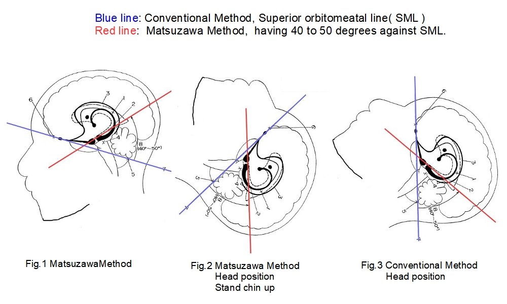
The OM reference line is used for general MRI, CT, and PET. It passes through the outer canthus of the eye and the center of the external auditory meatus. In the Matsuzawa method, MRI, CT, and PET are performed at a 40 to 50-degree angle to the OM reference line.
My understanding of the Matsuzawa method is simply by raising the chin. The wobble of the neck needs to be fixed with an appropriate jig. It is a pillow.
At a glance at the patent, it is not clear how the patented imaging method can be achieved by simply using a pillow. Most people may think that it requires modification of complex MRI, CT, and PET equipment. My interpretation of finding the realization with the pillow is also worthwhile.
If Dr. Matsuzawa’s patent can be realized with a single pillow, without the need to modify existing MRI, CT, or PET equipment, then everyone will adopt the imaging method when the patent expires.
The shape of the pillow needs to be slightly modified, but I believe that this can be adequately handled at each imaging site.
Comparison with Matsuzawa method and conventional imaging methods
will explain Dr. Matsuzawa’s photographing method in an easy-to-understand manner by slightly modifying the photo attached to the patent.The red line shows the photographic plane of Dr. Matsuzawa’s method.
The blue line is the conventional imaging plane and is the OM reference line. Strictly speaking, it is called the SM line, but it is described as the OM line in the patent document.
Figure 1 below shows a conceptual diagram of the cross-sectional method of imaging. Figure 3 shows the conventional standard imaging method, in which the OM reference line is perpendicular and the cross-sectional view is created when the patient is lying normally in the MRI or CT scanner.
Figure 2 shows an image taken using the Matsuzawa method. The angle of inclination is 40 to 50 degrees to the SM line. The optimal angle of inclination is determined at the time of imaging. This is the position where the tomogram of the hippocampus, amygdala, brain stem, and cerebral cortex see together.
The best angle is found within a range of 40 to 50 degrees, so whether the reference line is OM or SM does not affect the results.
It is a little tight, but you can raise your chin to create that imaging position. Notice the contour under the chin, which is a chin-up.
In this posture, they take several cross-sections (slices) with a thickness of 6 mm and a spacing of 2 mm, or with a thickness of 5 mm and a spacing of 1 mm.

Comparison with Matsuzawa method and conventional imaging methods
Problems with conventional imaging methods
Method 1
This is the case of Fig.3 above. This is an excerpt from the patent text.With this method, it is not possible to take tomographic images of the hippocampus and amygdala of the limbic system, the brainstem, and the cerebral cortex all on the same image plane.
In addition, it is difficult to obtain a complete picture of the limbic system because images of one part of the hippocampus and amygdala are taken in several cross-sections.
Furthermore, in many cases, it is difficult to even identify the hippocampus or amygdala in the images.
Method 2
This is another version of the conventional imaging method. The image is taken in the vertical section of the reference line in Fig. 3. The excerpt from the patent text has been slightly modified.Compared to conventional imaging method 1, this method produces images that are relatively easier to identify as hippocampus or amygdala.
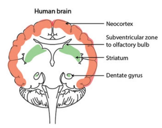
But as shown in Fig. 4, only a single image of a part of the hippocampus or amygdala is captured over several cross sections.
Fig. 4 below shows a cross-section perpendicular to the OM reference line. As pointed out in the patent, part of the hippocampus appears as a small, round outline in the cross-section. The entire hippocampus cannot be seen. DentateGyrus is the dentate gyrus of the hippocampus.

Fig.4 Cross-section perpendicular to the OM reference line,
From Sites of neurogenesis in the human brain
from SearchGate CC BY 4.0,
Figure adapted, with permission from Company of Biologists, from Magnusson and Frisen.
References
★Patent: Method and apparatus for producing tomographic images of the brain for close examination of the limbic system *1)Japan Patent Application 2002・43959, Application date: Nov. 26, 2001
★METHOD AND APPARATUS FOR TAKING CEREBRAL LAMINOGRAMS FOR WORKING UP LIMBIC SYSTEM *1)
International Patent: WO-A1・2/041775, Application date: Nov. 26, 2001